HUMAN CARDIAC AND RESPIRATORY SYSTEMS

Wednesday 12 May 2010

5.2 Relationship between diet, blood pressure, blood cholesterol and circulatory disease.

Our Diet is very important for our body systems and organs. The wrong kind of diet can cause many complications deep within the body that we are unaware of until something goes wrong.
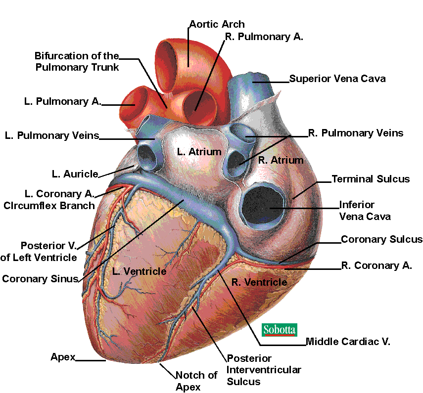
High blood pressure, high blood cholesterol and circulatory disease can all be linked with our diets.
If our food intake was high in saturated fats then the cholesterol levels in the blood would rise. This would cause a build up of fatty deposits sticking to the artery walls and forming atheromas.
High blood pressure means that the heart is beating harder than normal to try and push the blood around the body. The walls surrounding the blood vessels will eventually be damaged and so may some organs. It also increases the risk of angina attacks, heart attacks and strokes.
High blood pressure and blood cholesterol levels can be linked together. When the arteries become hardened by the cholesterol deposits the heart becomes more strained as it has to work even harder to pump the blood through the narrow passageways of the arteries in order to reach the organs.

So diet, blood pressure and blood cholesterol are all linked together and cause circulatory disease. There are easy steps to combat this disease,
These are;
  1. 30 minutes of exercise a day.
  2. reduce salt intake.
  3. limit alcohol intake.
  4. quit smoking
  5. lose excess body weight (if you are overweight)

1.2 Effective Gaseous exchange

Gaseous exchange occurs within the Alveoli in the lungs and the capillary walls.
Gaseous exchange means the diffusion of oxygen, carbon dioxide, water and other materials takes place. The oxygen diffuses through the membrane of the alveoli in to the capillaries and carbon dioxide diffuse from the capillaries into the alveoli where it is expelled from the body when expiring.
When Gaseous exchange occurs the gases only have to diffuse through two cells. These are the epithelial cell of the alveoli and the capillary wall.
In order to succeed with effective gaseous exchange a number of conditions must be met, these are large surface area and thin walls which allow the diffusion rate of the gases to be much faster,
if there is a lot of alveoli within the lungs then more gaseous exchange can occur (The alveoli can increase through exercise)
Water being diffused by surrounding calls keep the alveoli walls moist to allow effective gas exchange.
Faster diffusion rate between blood and alveoli will normally occur when the right temperature is met because it is more effective in warmer conditions.

Smoking Cessation - WHAT SMOKING DOES TO YOUR BODY

Here are the facts on smoking and the effects that it has on the body.
Smoking Cessation - WHAT SMOKING DOES TO YOUR BODY

The nervous system is affected because of the toxins inhaled from a cigerette disrupts the signals that the brain has sent to other body systems.
The respiratory systemhas reduced lung function and breathlessness because the lung airways become narrow and swell. The alveoli become permanently damaged and there is an increased risk of infections within lungs such as coughing and wheezing.
The circulatory system is affected by the tobacco smoke some of the effects are higher blood pressure and heart rate, blood becomes more prone to clotting, tightening of blood vessels, lower oxygen supply to organs and atherosclerosis.
Smokers are more prone to infections because the immune system doesn't function properly, there are lower levels of antioxidents in the blood and infections take longer to clear.
The musculorskeletal system is also affected becuse some of the muscles becomer tighter due to the toxins produced by the cigerette smoke and also bone density is reduced.

The relationship between smoking and Coronary Heart Disease (CHD)and lung cancer


Smoking is one of the factors that can trigger Coronary Heart Disease (CHD). The nicotine in a cigarette can raise blood pressure. It does this by constricting the blood vessels which will decrease the amount of blood supply to the vital organs. Nicotine can also increase the blood cholesterol levels which would normally only occur by a high fat diet.
Heart rate increases because the nervous system detects high levels of carbon monoxide in the blood. Carbon Monoxide enters the red blood cells and there is not enough room left in cell for oxygen so oxygen becomes restricted. The heart has to work harder to elliminate the toxins out of the body and supply enough oxygen to the heart and other organs. As the nervous system sends signals to the heart to make it work faster there is still not enough oxygen being received at the vital organs. The heart keeps pumping and soon it give up and cause Angina attacks or even worse a fatality.


a href="http://3.bp.blogspot.com/_MxP9XQgGOxY/S-sWp-hqFzI/AAAAAAAAADI/D4xaNWG7G28/s1600/lung+cancer.jpg">

"This is a chest x-ray of a person with bronchial cancer. This is a front view. The lungs are the two dark areas. The heart and other structures are white areas visible in the middle of the chest. The light areas that appear as subtle branches extending from the center into the lungs are cancerous.


Many people around the world suffer from lung cancer. The majority of it is related to smoking.
Many substances in a cigerette can cause damaging affects on the body and other substances make the cigerette taste nicer so its more addictive.
Cigerette smoke contains: Nicotine, Carbon Monoxide, Tar and other irritants..
Tar is the main substance that causes the cancer. The tar is made up of many different chemicals, Carcinogen is one of the main chemicals that can cause or aggravate cancer.

Sunday 9 May 2010

5.1 Describe the changes in artery structure associated with circulatory disease.

Heart disease is mainly caused by choice of lifestyle. The main offenders of this is smoking and diet.
When arteries become clogged this means that the heart will be receiving an insufficient supply of Oxygen which can lead to a condition known as Angina or even a heart attack.
Many different Arteries around the body could be subject to blockages and can affect many different organs and body parts.

picture from; http://www.nlm.nih.gov/medlineplus/ency/imagepages/18018.htm


Coronary Artery Disease (CAD) occurs when fatty deposits known as plaque build up in the artery walls allowing little or no blood to pass through. This forms a blood clot.
Too much cholesterol build up in the blood stream is one of the major causes of CAD. The plaque forms in the arteries over many years in a process called atherosclerosis. The plaque hardens in the passageways of the arteries which supply Oxygen rich blood to the major organs. Also pieces of the plaque fall of the wall of the arteries and travel in the blood stream where it can eventually block the flow of blood.


picture from:
http://www.nhlbi.nih.gov/health/dci/Diseases/Atherosclerosis/Atherosclerosis_WhatIs.html

The Arterial walls consists of the endothelium which is the inner wall that allows the blood to flow through smoothly, the media which is the muscle and elastic tissue that allows for the artery to cope with the high pressure of blood flowing through, and the adventita, this is the tough outer shell that protects the artery.
Carotid artery disease is when the arteries in both sides of the neck become blocked due to the fatty deposits clogging up on the endothelium. This causes the brain to have an insufficient supply of oxygen rich blood and can lead to stroke.
Pheripheral artery disease occurs when major arteries in the legs, arms and pelvis become blocked due to the fatt deposits. This can lead to serious infections (gangrene) or unbearable pain.
High blood pressure can damage the vessel walls because of the blood being constantly forced through them.
Some of the circulatory diseases may occur because they are heriditary i.e. high blood pressure,
These conditions can be avoided.

Redistribution of blood

When exercising the circulatory system adapts, this is because the need for more oxygen entering the muscles is important. The majority of blood during exercise flows mainly to the heart, the brain and the working muscles. The vital organs have minimal blood flow.
Arteries and Veins can either constrict or dilate during exercise as this is the most efficient way of redistributing blood. In the working muscles the arteries dilate so the increase of blood can flow through the capilleries more effectively. This means that there is an increase of Oxygen supplied and a decrease of carbon dioxide in the muscles.
The nervous system also adapts as it secretes hormones which signals the body when exercise begins. The hormones signal the the blood vessells to dilate in the heart and the working muscles.

Saturday 8 May 2010

The role of the nervous system in generating normal breathing rhythm

The heart is able to contract on its own accord but it will be unable to keep a constant rhythm of beating. This is where the Nervous system comes into effect. Impulses are sent from the brain in order to control the speeding up of the heart rate and the slowing down of the heart rate depending on it's need at a particular time.


The nervous system is critical for our every day living and functioning of the body. The Brain is divided into 3 parts, these are the Cerebrum, the Cerebellum and the brain stem. The central controlling area for breathing is situated in the lower part of the Brain stem known as the Medulla Oblongata. Also situated in the Brain stem is the Pons and the Midbrain.
It is the Brain stem along with the spinal cord and medulla oblongata that controls the autonomic functions and the transporting of nerve signals between between brain and spinal cord.(The spinal cord serves as a pathway for the nerve impulses as they travel to and from the brain.)
The autonomic nervous system sends impulses to the vital organs within our body. It functions without conscious effort and controls the heart rate, breathing rate, blood pressure and body temperature.


There are different types of neurons in the body, inspiratory neurons and expiratory neurons are responsible to maintain the normal breathing rhythm. The inspiratory neurons are active when inspiring and inactive when expiring and the expiratory neurons are active when expiring and inactive when inspiring. The neurons are sent to the diaphragm and intercostal muscles with messages signalling them to contract and relax at regular intervals.

There are two different nerve cells that detect the levels of change in oxygen and carbon dioxide. These are pheripheral chemoreceptors and central receptors. The pheripheral chemoreceptors monitor the levels of oxygen in the blood, if the oxygen decreases then they send messages to the respiratory centre to increase the rate and depth of breathing. Carbon dioxide is censored by both receptors. The central receptor is situated in the medulla. It monitors the levels of carbon dioxide in the cerebrospinal fluid. This fluid surrounds the brain and the spinal cord. If the carbon dioxide levels are too high then the receptors are triggered, they send impulses to the respiratory centre to increase the breathing rate allowing more oxygen into the blood and allowing the carbondioxide to diffuse into the capilleries so that it can exit the body.

Wednesday 28 April 2010

The cardiac cycle


We all know that the main purpose of the heart is to pump blood around the body so that our vital organs and our muscles can receive the oxygen that they require!!

In this part I will be looking at the cardiac cycle of the heart and how this process works!

The heart is a cardiac muscle it has four chambers these are left ventricle, left atrium, right ventricle and right atrium.(The atrium's are at the top of the heart and the ventricles are at the bottom of the heart)
Oxygenated blood passes through the pulmonary vein in the left atrium, when the atrium is full the atrioventricular valves open allowing the blood to be pushed into the ventricles. As the blood enters the ventricles the atrioventricular valves stop the blood from flowing back into the atrium. The ventricles contract pushing the blood through the semi-lunar valves into the aorta. This is where the blood leaves the heart and travels around the body via the arteries, arterioles and capillaries. Deoxygenated blood enters the heart through the superior vena cava and the inferior vena cava into the right atrium, as the pressure builds up in the atrium the semi-lunar valves are pushed open allowing the blood to enter the right ventricles. When the heart contracts the deoxygenated blood is forced into the pulmonary artery so that the blood can enter the lungs and become oxygenated.

Did you know that your heart beats 70 times a minute! and that each heartbeat lasts around 0.8seconds!

Within each heart beat the heart relaxes and contracts this is known as systole(contract) and diastole (relax). The heart is relaxed longer then it is contracted. This is because the nervous system which is in a part of the brain called the medulla oblongata sends electrical impulses to the heart in order to stimulate the sinoatrial node (SAN), This is know as the hearts natural pacemaker. These electrical impulses allow the cardiac muscle to contract the right and left atrium allowing the blood to pass through the atrioventricular valves into the relaxed ventricles. There is a slight delay here so that the atria can empty fully. The electrical impulses spread into tissues called Bundle of His. These tissues connect the atrioventricular node to the septum. The bundle of his has branches leading into the ventricle walls known as purkinje fibres. The purkinje fibres allow the impulse to travel through them so that they can get to the ventricle walls. as this happens it the impulses cause the ventricles to contract (also known as ventricular systole). The impulses are stronger at the ventricles because the blood has further to travel than the atria.
This process is repeated every time the heart beats and it is known as THE CARDIAC CYCLE.

Cardiac output is the volume of blood pumped by each ventricle in one minute.
It is calculated by a simple sum Q = SV x HR (this means Q(cardiac output) = SV(stroke volume) x HR(heart rate)
Measuring Cardiac output is extremely important as it allows us to know how healthy a heart is. This allows us to know if the heart is under strain i.e. heart failure. In a healthy human being the average cardiac output is around 5 litres of blood per minute.

The heart has its own system called a pacemaker which is connected to the SA node located at the top of the heart. It contains sympathetic and parasympathetic nerve fibres. These fibres release hormones which can either slow the heart rate down or speed the heart rate up. These hormones are delivered under conditions of rest(parasympathetic nerve fibres) or emotional and physical activity (sympathetic nerve fibres).

Blood plasma


Plasma is the liquid part of the blood, it transports dissolved substances around the body. Also it fights against disease and removes unwanted waste materials from cells.
the blood plasma helps to maintain body temperature and control the pH of blood. Another function of plasma is that it contains clotting agents so when blood is exposed to air it will clot. this is to prevent anymore bacteria entering the body.
Plasma contains 90% water and the other 10% consists of glucose, amino acids, vitamins, mineral salts, hormones, antibodies and antitoxins. all of the different substances in the plasma have a job to do. for example; amino acids and glucose travel in the blood so that they can be diffused into areas that need it i.e. if there is muscle tissue damage then the amino acids help to repair it and they help with growth. if there is no damage that needs repairing then the substances will be stored and used later when they are need for anaerobic respiration. the glucose will travel to muscles for storage or they will travel to the liver where the hormones which travel in the plasma will help to turn glucose into glycogen.
Antibodies and antitoxins travel in the blood plasma as this is the most efficient way for them to get to areas of infection quickly.


http://www.ivy-rose.co.uk/HumanBody/Blood/Blood_StructureandFunctions.php

Tuesday 27 April 2010


The structure and function of Red Blood Cells
Red blood cells (RBC) are produced in our bones. our bones reproduce them constantly because red blood cells only have a life span of 120days. The cells become worn out and die. They are the primary cells in our body as their function is too deliver oxygen to our cells and remove carbon dioxide and other waste materials.
RBC have biconcave discs this means that the centre of both sides of the cell is depressed. The depressed centre allows the cells to have more cell membrane surface to allow the diffusing of oxygen and carbon dioxide to take place. This shape also allows for the RBC to have a large surface area for oxygen. Due to the structure of the RBC they are more flexible than other cells and they can squeeze through tight passages such as capillaries between cells in tissues. They can also bend and return to their original shape.
RBC contain a protein chemical called hemoglobin. This protein gives the cells their 'red' colour. Hemoglobin also contains iron. When a red blood cell is first produced it has a nuclei. As it matures it loses the nuclei, this is to allow more space for the haemoglobin so that it can carry as much oxygen as possible. As the RBC passes through the lungs oxygen molecules attach themselves to the hemoglobin. The blood travels around the body and allows the hemoglobin to release the oxygen into cells . once releasing the oxygen the hemoglobin has a large surface area that it needs to fill so it bonds with carbon dioxide and other waste products and transports them back to the lugs so that they can be eliminated from the body. This cycle continues constantly.

Heart Anatomy

video on the anatomy of the heart!


Tuesday 20 April 2010

Blood

Blood has two components, plasma and cells. Plasma is the liquid part of the blood. it is made up of 90% water and the other 10% are materials that are essential for life. i.e. nutrients and vitamins - these are dissolved in the blood plasma.
The cell part of the blood contains RBC (Red blood cells), WBC (White blood cells) and platelets. The RBC consists of a protein called Hemoglobin which carries oxygen from the lungs around the body, they also help with the removal of carbon dioxide. When RBC are produced they contain a nucleus as the RBC matures the nucleus leaves the cell. The WBC fight infections, they are often referred to as Leukocytes. The WBC are part of the immune system and they circulate in the blood stream so that they can be transported to infected areas in the body. The platelets are used by the body for clotting.
as the blood circulates the body the materials that are in the plasma diffuse into cells and tissues. they diffuse from high concentrated areas too low concentrated areas (in other words the higher the difference in concentration the higher the amount of materials diffused.)


This is a picture to show what the platelets do when they begin to clot.

Monday 19 April 2010

structure of arteries, veins and capillaries

An artery is a vessell that carries blood away from the heart. All arteries except for the pulmonary artery and its branches carry oxygenated blood. small arteries are called arterioles. Arteries have elastic tissue in their wall and they have a thick muscle layer in the walls aswell. This structure allows the arteries to cope with the high pressure of blood flow caused by the heart beat. The endothelium is folded allowing the artery to expand.


A vein carries blood towards the heart. All veins except for the pulmonary veins contain deoxygenated blood small veins are called venules. the main veins of the body have valves to prevent blood flowing in the wrong direction. Contraction of body muscles help the blood flow through the veins. Veins are wider than arteries and they have very little elastic or muscle tissue. The space in the centre of the vein is much larger than the arteies.


Capillaries are vessells which transport the blood from arterioles to venules. they are microscopic vessells which are in most of our ogans and tissues in the body. The structure of the capillaeies are very important because it helps with the function of what it does. They are only one cell thick so that they can allow the exchange of oxygen, carbon dioxide, minerals, salts and water to occur between the capillary and the surrounding tissues.

Structure of the respiratory system

The organs of the respiratory system consists of the nose, pharynx, larynx, oesophagus, trachea, bronchi and the lungs.
The nose consists of three nasal cavities in order to supply the lungs with as mush oxygen as possible. the nose filters the air of impurities as well as warming and moistening the air before it enters the lungs.

The pharynx is a tube like structure which allows both air and food too pass through before they reach the appropriate tubes.
The larynx is situated below the pharynx and just above the opening of the trachea, it is part of the vital airway to the lungs.
the epiglottis is one of the main cartilages in the larynx it is small and is attached to the thyroid cartilage along one edge. the epiglottis protects the airway against solids and liquids entering during swallowing.
the trachea is a long tube which holds open the passage way so that air can reach the lungs from the outside. if this airway becomes obstructed for even a few minutes then it Will result in loss of consciousness because of the lack of oxygen supplied.
At the end of the trachea the tube divides into two parts known as the bronchi, each bronchi enter the lungs and branch of into bronchioles. Each bronchiole has little air sacs at the end known as alveoli so that gaseous exchange can take place. Gaseous exchange is when oxygen is diffused into the blood capillaries from the alveoli and carbon dioxide is diffused into the alveoli from the blood capillaries.


http://www.facebook.com/l.php?u=http%3A%2F%2Fwww.ambulancetechnicianstudy.co.uk%2Frespsystem.html&h=d2b8575ab4255a1d6fad5e70a90c4925